In this video Dr. Learn MRI Report Reading.

Healthcare Extreme How To Read An Mri Lumbar Spine In 8 Easy Steps
MRI - Slipped Disc - Book Appointments Zocdoc can help you find top-rated doctors who can perform an MRI for a slipped disc.

. How to Read Report of MRI. Varun Wasil- MPTOrthopaedics from Sukoon Physical Therapy Jalandhar. MRI of the cervical spine was obtained with the following sequences.
T1 weighted sagittal and coronal T2 weighted coronal FLAIR and T1 weighted axial sequences. This is the 1-3. An MRI spine is a unique test that tells us about your spinal cord discs and bones.
Axial GRE sagittal T1 T2 and STIR. Learn to Read your MRI Report Lumbar spine MRI Reading Easy MethodIn this video Dr. Also due to the fact that an MRI is able to focus on a.
Its simple secure and free. About Press Copyright Contact us Creators Advertise Developers Terms Privacy Policy Safety How YouTube works Test new features Press Copyright Contact us Creators. A bulging or herniated disc.
A total 109 patients of lumbar disc degeneration were diagnosed on 15 Tesla MRI machine. Mentioned below are some of the symptoms of slipped disc. MRI scans are often used to diagnose and monitor herniated discs.
MRI scan confirms the location and severity of the slipped disc and helps in finding details of the spinal infection or presence of a possible tumor. Disc bulgesherniations are when there are damage to these shock absorbers that can lead to pressure on the spinal cord or nerves that come off of it. An MRI scan has a number of distinct advantages that benefit a herniated disc patient including that it is unobtrusive painless and free of radiation.
Worsening of the pain especially during night or with. The one thing after an MRI is the MRI radiology report. Pain extending to arms and legs.
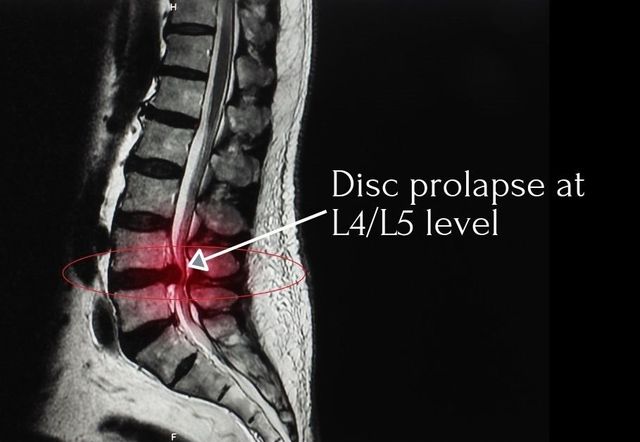
Disc prolapse occurs commonly in middle age with a typical history of an episode of back pain either related to lifting and or twisting or which occurs spontaneously. MRI examination of the brain is performed using the following protocol. Magnetic Resonance Imaging is an amazing tool that allows us to see deep inside the human body with a degree of clarity that is absolutely amazing.
MRI scans as a medical tool. We get a good view of the spinal canal bony vertebrae and disc spaces between vertebrae spaces contain fluid nerve. Varun Wasil from Sukoon Physical Therapy Jalandhar Punjab explained about MRI report.
Diffuse disk bulge are seen at l3 -4 and l4-5 levels with indentation of the these sac and obliteration of corresponding lateral recesses. One of the most common ways they are used is to identify the location of the herniated disc s in the spine and the degree of nerve. It helps in detecting any tear herniation or fragmentation of.
We can visualize the tiny. Primary Care Physicians Browse. All the observation was done by three Radiologists Professor Associate Professor and.
Numbness and pain mostly on one side of the body. You may also have heard the term slipped disc. 80 of disc prolapses.
Maybe a lot of physical therapy. There is straightening of the normal cervical lordosis which may be positional.

How To Read Mri Report Of Slipped Disc L4 L5 L5 S1 Sipped Disc Diagnosis In Hindi Youtube

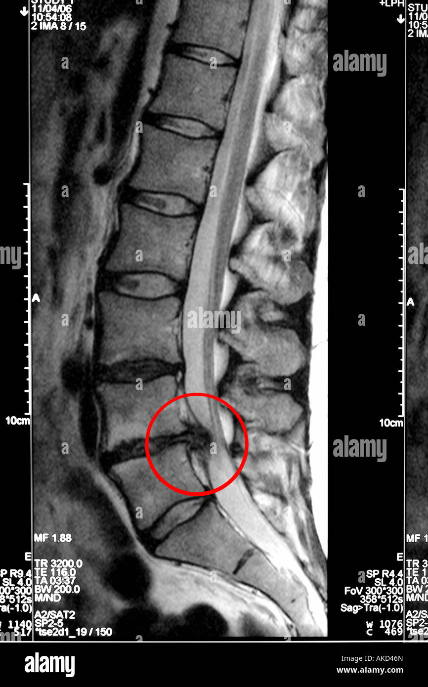
Mri Scan Clearly Showing A Slipped Disc Pressing On The Spinal Cord With The Affected Area Circled In Red Stock Photo Alamy

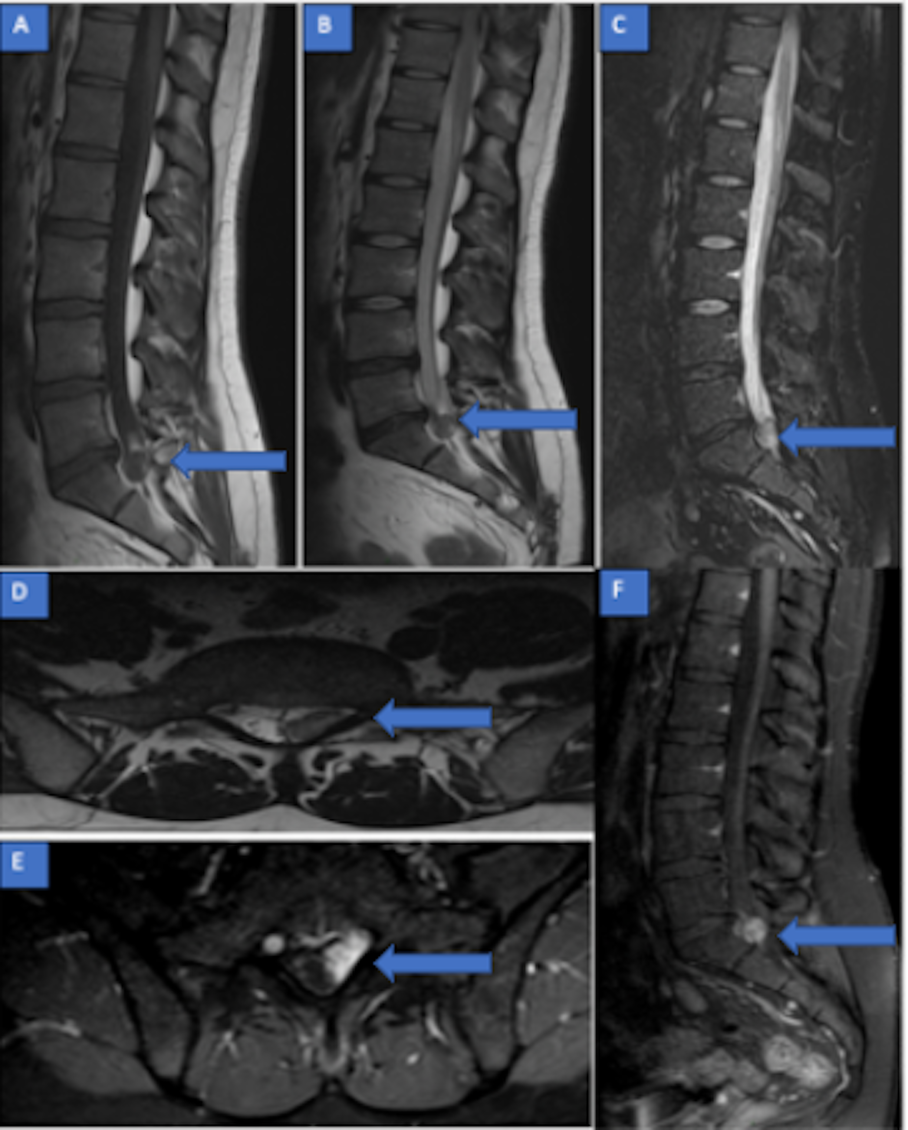
Lumbar Spinal Mri Of Patient 2 Demonstrating The L5 S1 Discal Cyst Download Scientific Diagram

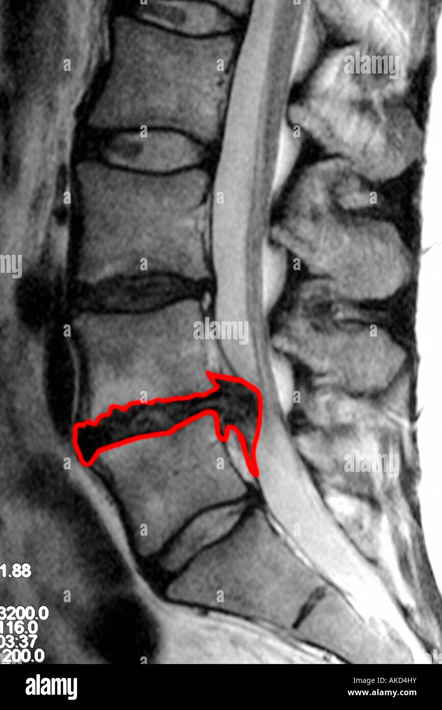
Mri Scan Clearly Showing A Slipped Disc Pressing On The Spinal Cord With The Affected Area Outlined In Red Stock Photo Alamy

What The Doctor Is Looking For In A Spine Mri Advanced Bone Joint

Healthcare Extreme How To Read An Mri Lumbar Spine In 8 Easy Steps

How To Read A Mri Of A Lumbar Herniated Disc Lower Back Pain Colorado Spine Surgeon Youtube

Magnetic Resonance Imaging Or Mri Scan Report Of Spinal Cord Or Lumbar Bulging X Ray Lumbar Spine Low Back Lower Disc Bone Stock Image Image Of Medical Exam 238804081

Axial Ct Scan A Axial B And Sagittal C Mri On T2 Weighted Download Scientific Diagram

T2w Mri Showing A Huge L4 L5 Herniated Disc Download Scientific Diagram

Cureus Mri Of Acute Low Back Pain About An Uncommon Pitfall

Mri Scan Left C6 C7 Herniated Disc Figure 5 Mri Scan Left C6 C7 Download Scientific Diagram

Lumbar Disc Herniation Mri Explained Dr Jeffrey P Johnson Hd Youtube

Slipped Disc Mri Scan Stock Image M330 0976 Science Photo Library
How To Read The Mri For A Herniated Disc And 5 Treatment Options

Mri Documentation Of Spontaneous Regression Of Lumbar Disc Herniation A Case Report Semantic Scholar

Slipped Disc Mri Scan Stock Image M330 1622 Science Photo Library

Lower Back Pain The Herniated Disc
